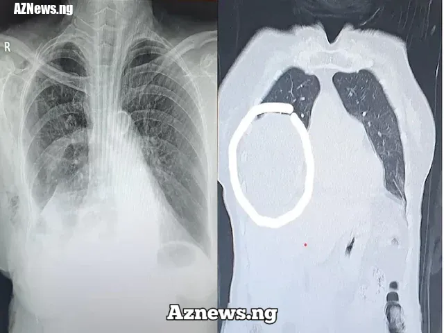
The woman was suffering from shortness of breath for over an year; the tumour in the right side of her chest had compressed her right lung, compromising her lung capacity......Read The Full Article>>.....Read The Full Article>>
A team of doctors in Hyderabad removed a big tumour from the chest of a 69-year-old woman who was struggling with breathing difficulties for the past one year. The rare pleural tumour (18 x 15 cm size) was found in the right side of her chest, arising from the lining of the chest wall.
According to the doctors at Yashoda Hospitals, where the woman was operated, the heart was pushed to the opposite side due to the tumour, thereby compressing the right lung and the diaphragm. This had resulted in fluid accumulation in her right chest, causing significant chest pain.
The woman was suffering from shortness of breath for over an year, said Dr. Balasubramoniam KR, Consultant Minimally Invasive and Robotic Thoracic Surgeon at Yashoda Hospitals, while talking about the case.
Owing to severe compression of the right lung, which had weakened her lung capacity, she was experiencing poor pulmonary function. Still, she was operated as immediate removal of the tumour was required to avoid further compression of the lung, spine, heart and blood vessels, and prevent deadly complications, Dr. Balasubramoniam stated.
Watch out for the persistent nonresolving symptoms
“Chest cavity is quite capacious enough to allow tumours to grow to massive sizes,” said Dr. Balasubramoniam, warning people to be aware of the persistent nonresolving symptoms.
“Some tumours grow to massive sizes and pose challenges in surgery and anesthesia but can be removed safely if the patient is evaluated and assessed properly before surgery,” he added.
In this case, the woman was discharged three days after surgery, free of respiratory symptoms.
Hence, doctors encourage people to go for routine health checkups, especially if they have persistent unresolving symptoms, even for minor symptoms. Tumors in the chest can be easily evaluated with routine tests like chest X ray and CT.
Remember, early detection is the key to lesser surgical risk and earlier recovery. Also, proper presurgical evaluation and assessment improves surgical results and recovery
